Digestive System Organs
MCAT Biology - Chapter 7 - Section 6.2 - Organ Systems - Digestive System
- Home
- »
- MCAT Masterclass
- »
- Biological and Biochemical Foundations of Living Systems
- »
- Biology
- »
- Digestive System Organs – MCAT Biology
Sample MCAT Question - Digestive System Organs
Secretion of gastrin is inhibited by:
a) High HCl levels
b) Stomach distension
c) High calcium levels
d) Vagal stimulation
A is correct. High HCl levels. The release of gastrin can be inhibited in specific ways, one of which is the presence of acid (HCl) in the stomach. One of the main functions of gastrin is to stimulate parietal cells to release HCl into the stomach, so by negative feedback, the presence of HCl would inhibit gastrin secretion. Answer choices B, C and D all stimulate gastrin release.
Get 1-on-1 MCAT Tutoring From a Specialist
With MCAT tutoring from MedSchoolCoach, we are committed to help you prepare, excel, and optimize your ideal score on the MCAT exam.
For each student we work with, we learn about their learning style, content knowledge, and goals. We match them with the most suitable tutor and conduct online sessions that make them feel as if they are in the classroom. Each session is recorded, plus with access to whiteboard notes. We focus on high-yield topics if you’re pressed for time. If you have more time or high-score goals, we meticulously cover the entire MCAT syllabus.
Digestive System Organs
In this MCAT content post, we’ll discuss several crucial organs of the digestive system, including the mouth, esophagus, stomach, small intestine (duodenum, jejunum, and ileum), and finally the large intestine. By examining each of these organs and their function, we’ll get a better idea of how the entire process of digestion works.
Mouth
The mouth is the first part of the digestive system and is where the ingestion of food occurs. The first process of ingestion is chewing. Chewing is a mechanical process that helps to break down food into smaller particles. It also mixes the food with saliva, which is essential for lubricating the food to facilitate swallowing. If food were entirely dry, it would be tough to swallow it.
Moreover, saliva also contains several enzymes that aid in the chemical breakdown of food into nutrients. Salivary amylase breaks down carbohydrates such as starch into smaller carbohydrates that can be digested. Salivary lipase is another salivary enzyme that breaks down triglycerides, or fats, into free fatty acids. Lastly, lysozyme is an enzyme that’s found in saliva, and also several other places such as tears and even human breast milk. It is part of the innate immune system and functions to kill bacteria by breaking down the peptidoglycan cell wall.
Esophagus
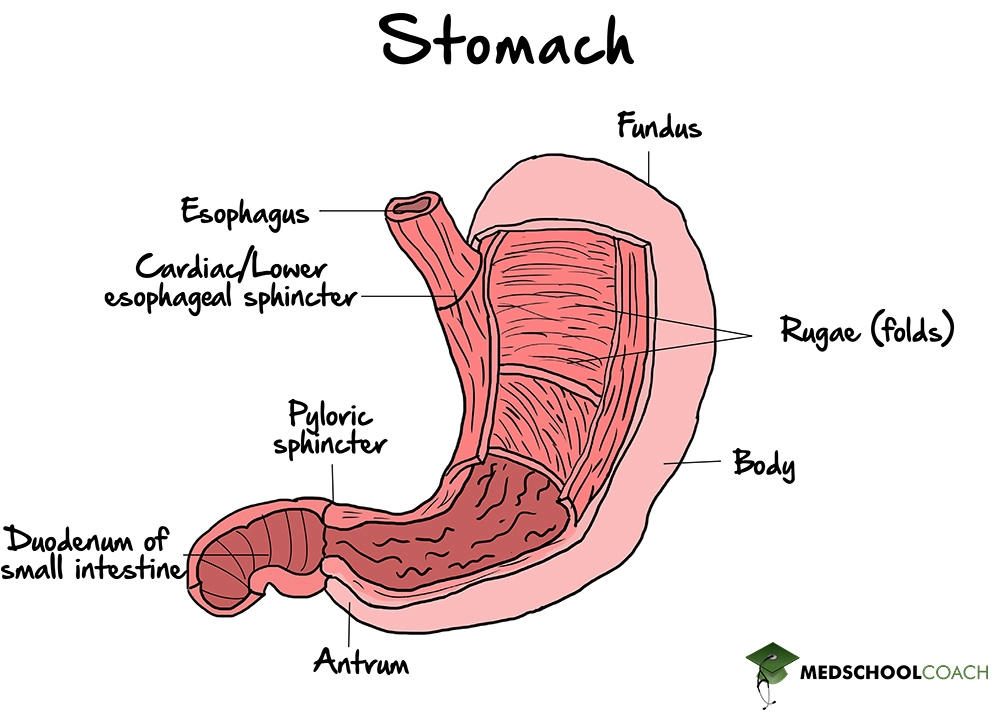
From the mouth, food moves into the esophagus, which is a tube that connects the mouth to the stomach. Peristaltic contractions move food along the esophagus to the lower esophageal sphincter, also known as the cardiac sphincter or gastroesophageal sphincter, which opens to allow food to enter the stomach. Figure 1 shows an image of the stomach with the cardiac sphincter. Aside from letting food into the stomach, the sphincter also prevents food from reentering the esophagus. If this happens, it can lead to what is known as heartburn, or gastroesophageal reflux disease (GERD). GERD can be a severe problem because if the acid from the stomach is allowed to reflux into the esophagus, it can lead to esophageal irritation, inflammation, and over time, it may even lead to cancer.

Stomach
The stomach is necessary not only for digestion but also for the storage of food. When food enters the stomach, the stomach distends. This distension causes G cells in the stomach to release gastrin. Gastrin is a peptide hormone that acts on parietal cells in the stomach to release hydrochloric acid (HCl). The stomach needs to have a low pH in order to carry out its digestive functions. However, a pH that is too low can cause gastric ulcers. In this way, gastrin secretion is inhibited by the presence of acid or a low pH in the stomach, which is an example of negative feedback.
Furthermore, HCl in the stomach acts on pepsinogen, which is a zymogen. Pepsinogen is produced by the chief cells that line the stomach mucosa. In the presence of HCl, pepsinogen is converted into its active form, pepsin, which is a protease that breaks down proteins into smaller amino acids for digestion. Finally, food exits the stomach through the pyloric sphincter (Figure 1). The pyloric sphincter is the junction between the stomach and the small intestine, and it has a role in regulating the release of chyme into the small intestine. Chyme is the term for food particles and digestive juices from the stomach. Its release into the small intestine has to be carefully regulated to allow for optimal absorption of nutrients.
Small Intestine
The small intestine can be broken down into three segments – the duodenum, the jejunum, and the ileum. The duodenum connects directly to the stomach, the jejunum connects the duodenum to the ileum, and the ileum is connected to the large intestine. All three segments are involved in digestion and absorption of nutrients.
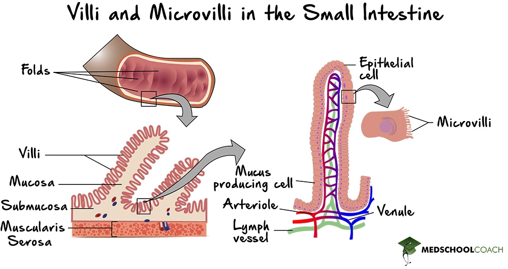
Figure 1 details the surface area of the small intestines, and details how the small intestine can absorb large amounts of nutrients. The intestinal epithelium is folded, and the folds contain villi, which are protrusions of the epithelium. Within the villi, the individual epithelial cells contain microvilli. The purpose of the villi and microvilli is to massively increase the surface area of the small intestine in order to increase the rate of nutrient absorption.

In terms of digestion, when the pyloric sphincter relaxes, chyme is released into the duodenum. The presence of chyme in the duodenum causes the duodenum to secrete the hormones cholecystokinin (CCK), as well as secretin.
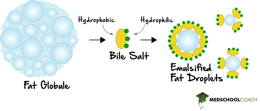
CCK acts on the gallbladder to secrete bile, which has a vital role in emulsifying fats (Figure 2). Bile is a salt and is an amphipathic molecule, meaning that it has both hydrophilic and lipophilic properties. In this way, bile will aggregate towards large fat globules and can break them down into smaller fat globules, which significantly increases the rate at which fats can be broken down by the body. In the absence of bile, the body is unable to digest fat, and they are excreted in the feces. This condition is known as steatorrhea, and it can lead to fatty acid and vitamin deficiencies.

Another function of CCK is to stimulate the pancreas to release digestive enzymes. For each of the four biological macromolecules – carbohydrates, nucleic acids, proteins, and fats – there are specific digestive enzymes that break them down. Amylase breaks down carbohydrates, nuclease breaks down nucleic acids, protease breaks down proteins, and lipase breaks down fats.
Secretin, another compound released by the duodenum, stimulates the pancreas to secrete bicarbonate, which aids in digestion. Many digestive enzymes require a higher pH in order to function optimally. The chyme from the stomach, which enters into the small intestine, is at a low pH. Bicarbonate helps to increase the pH of the stomach contents in the small intestine to aid digestive enzymes.
The duodenum also secretes enterokinase. Enterokinase converts trypsinogen into trypsin. Trypsinogen is one of the digestive enzymes released by the pancreas. Many of these digestive enzymes are released as zymogens or in an inactive form. Enterokinase converts trypsinogen into its active form, trypsin. Trypsin will then aid in converting other digestive enzymes into their active forms.
Large Intestine
The primary role of the large intestine is to absorb water. People with diseases that cause excess loss of fluid through the gastrointestinal tract, such as diarrhea, generally have a problem with their large intestines. Also, the large intestine stores fat for defecation and also contains healthy flora beneficial for humans. Normal flora are the gut bacteria in all human GI tracts. These bacteria are good for human health and have formed a symbiotic relationship with humans. They can produce vitamin K as well as other essential nutrients for the body.
At the very end of the large intestine is the anus. The anus has both an internal and external sphincter. This anatomy is similar to the urinary sphincters in the excretory system. The internal anal sphincter is made of smooth muscle and is involuntary, and the external anal sphincter is made of skeletal muscle and is voluntary.
Explore More MCAT Masterclass Chapters
Take a closer look at our entire MCAT Masterclass or explore our Biochemistry lessons below.

One-on-One Tutoring
Are you ready to take your MCAT performance to a whole new level? Work with our 99th-percentile MCAT tutors to boost your score by 12 points or more!
See if MCAT Tutoring can help me
Talk to our enrollment team about MCAT Tutoring

MCAT Go Audio Course
Engaging audio learning to take your MCAT learning on the go, any time, any where. You'll be on the way to a higher MCAT score no matter where you are. Listen to over 200+ lessons.

MCAT Practice Exams
Practice makes perfect! Our mock exams coupled with thorough explanations and in-depth analytics help students understand exactly where they stand.

MCAT Prep App
Access hundreds of MCAT videos to help you study and raise your exam score. Augment your learning with expert-created flashcards and a question banks.
